- Undergraduate
- Graduate
- Research
- Community
- News & Events
- People
Back to Top Nav
Back to Top Nav
Back to Top Nav
Back to Top Nav
August 29,2018 by Joseph Blumberg
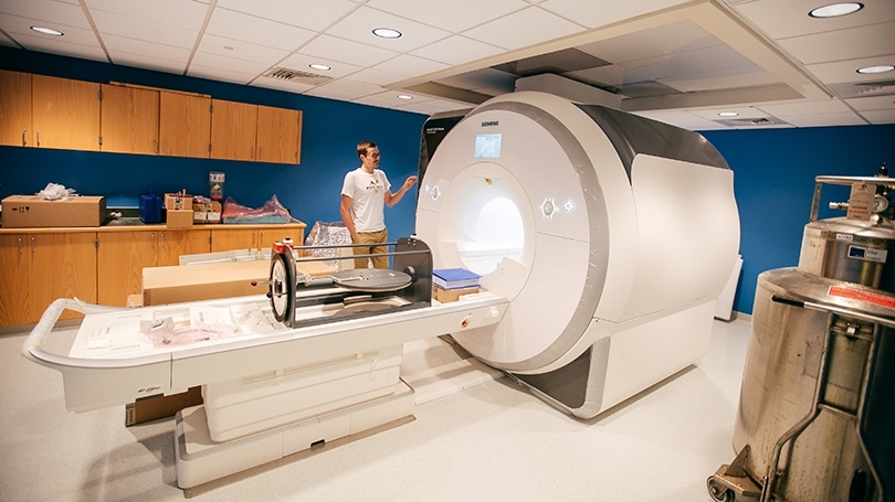
Researchers are welcoming the arrival of a new fMRI scanner, the latest in a series of scanners dating back to 1999, when the Dartmouth became the first liberal arts college in the nation to own and operate a functional magnetic resonance imaging device strictly for research purposes.
The new scanner, weighing more than 26,000 pounds, was lowered into its bay beneath Moore Hall last month, in the home of the Department of Psychological and Brain Sciences (PBS).
“This is a big deal,” says James Haxby, the Evans Family Distinguished Professor and director of both the Center for Cognitive Neuroscience and the Brain Imaging Center. “We are extremely excited about getting this new scanner. It will be in use seven days a week, from early morning to late at night.”
Over the years, the Brain Imaging Center’s fMRI scanners have been used for researching questions about the organization of the brain, the concept of free will, the role of the amygdala in learning and anxiety disorders, and how the brain recognizes music, visual imagery, and its relation to memory.
“Having an in-house MRI scanner has allowed us speed and flexibility in asking questions about how the mind is realized in the brain's neural activity,” says Professor of Psychological and Brain Sciences Peter Tse. “This has probably made my lab twice as productive as it would have otherwise been. It also affords undergraduates opportunities to get involved in cutting-edge research right here on campus.”
Todd Heatherton, the Lincoln Filene Professor of Human Relations and director of Dartmouth’s Center for Social Brain Sciences, notes that the fMRI has allowed researchers to identify an area in the brain associated with knowledge about the self.
“One of the most cited papers in cognitive neuroscience was our study showing the medial prefrontal cortex is involved in self-reflection,” says Heatherton. “It has been cited more than 1,300 times.”
An fMRI uses a strong magnetic field and radio waves to create detailed images of the brain. It looks at changes in blood flow in the brain to map areas of neural activity, providing potential insight into how the brain works.
“It is a tool we can use to decode brain activity and help us understand circuits for perception and circuits for decision-making,” says Haxby. “We can get much better answers, much clearer, more penetrating answers with better equipment.”
Noting that his department offers an undergraduate course utilizing fMRI, Haxby says, “We have a very popular course on fMRI imaging for undergraduates and several senior theses every year are based on assignments undergraduate have run on the scanner.”
Supported by funding from the National Science Foundation, the National Institutes of Health, and private foundations, the new scanner will serve as the principal research instrument for more than 15 faculty investigators, their graduate students and postdocs, and undergraduates.